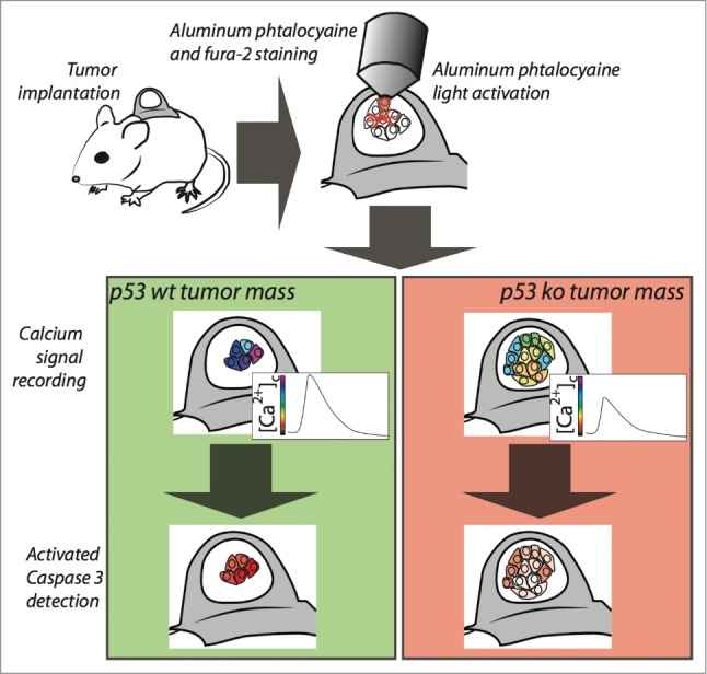
Figure 1.

Imaging of calcium signaling and apoptosis into tumor mass. Tumors grown within the skinfold chamber are loaded with aluminum phtalocyanine and the Ca2+ indicator Fura-2. Phtalocyanine activation and Ca2+ signal recording are performed in the same situ using the microscope optics. After Ca2+ live imaging, apoptosis is measured by intravenous administration of a fluorescent marker measuring caspase activity.
