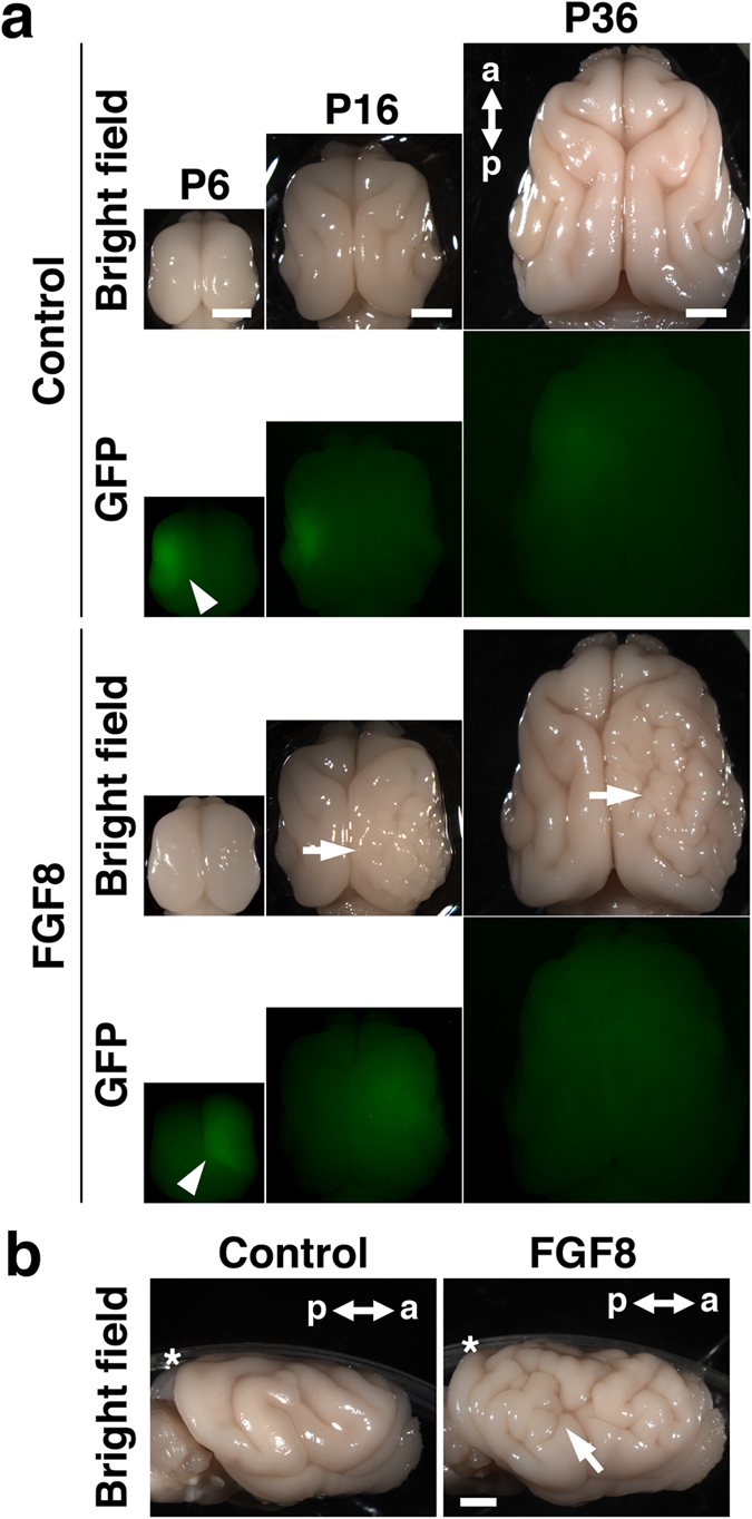
Figure 1. FGF8 induces polymicrogyria and megalencephaly in the developing ferret brain.

(a) Dorsal views of the developing ferret brain. GFP or GFP plus FGF8 were expressed in one side of the brain at E33 using in utero electroporation, and the brain was prepared at the indicated time points. Electroporated areas showed GFP fluorescence (arrowheads). FGF8 induced polymicrogyria and megalencephaly at P16 and P36 (arrows). (b) Lateral views of the ferret brain at P36. The arrow indicates polymicrogyria. Asterisks indicate plastic dishes. a, anterior; p, posterior. Scale bars = 4 mm.
