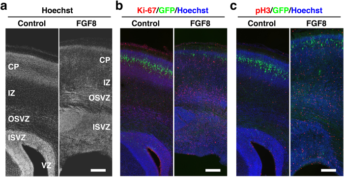
Figure 4. Cell proliferation in the cerebral cortex of developing TD ferrets.
GFP and FGF8 were expressed in the ferret cerebral cortex at E33 using in utero electroporation, and the brain was prepared at P6. Coronal sections were stained with Hoechst 33342 (white in (a), blue in (b, c)) plus either anti-Ki-67 antibody or anti-phospho-histone H3 antibody (red). Cortical regions containing transfected GFP-positive areas (green) are shown. Note that Ki-67-positive cells and pH3-positive cells were markedly increased in TD ferrets. Scale bars = 300 μm.

