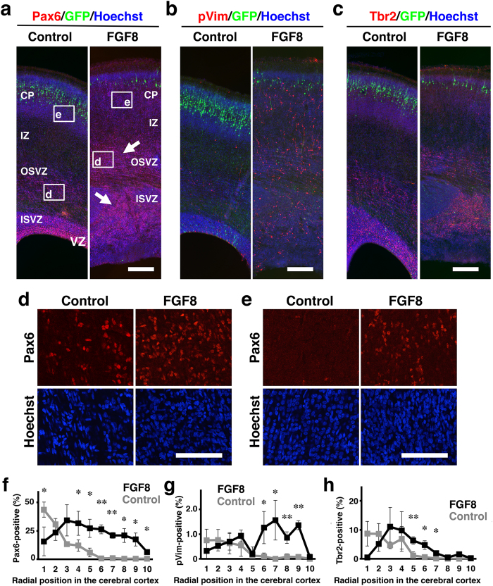
Figure 6. The distribution of Pax6-, pVim- and Tbr2-positive cells in the cerebral cortex of developing TD ferrets.
GFP and FGF8 were expressed in the ferret cerebral cortex at E33 using in utero electroporation, and the brain was prepared at P6. Coronal sections were stained with Hoechst 33342 (blue) plus either anti-Pax6 antibody, anti-phosphorylated vimentin (pVim) antibody or anti-Tbr2 antibody (red). (a–c) The cerebral cortex containing the transfected GFP-positive area (green) is shown. Note that Pax6-positive cells (arrows), pVim-positive cells and Tbr2-positive cells were markedly increased in TD ferrets. (d) Pax6-positive cells in the OSVZ. Confocal images in the white boxes in (a) are shown. (e) Pax6-positive cells in the CP. Confocal images in the white boxes in (a) are shown. Note that Pax6-positive cells were markedly increased both in the OSVZ and in the CP. (f–h) The cerebral cortex was divided into 10 regions along the radial axis from the ventricular surface (1) to the pial surface (10). The numbers of Pax6-, pVim- and Tbr2-positive cells in each region were counted and were divided by the numbers of Hoechst 33342-positive cells in the same region. The percentages of positive cells are shown. Bars represent mean ± SD. *p < 0.05; **p < 0.01. n = 3 for each condition. Scale bars = 300 μm (a–c), 100 μm (d,e).

