
Figure 2.
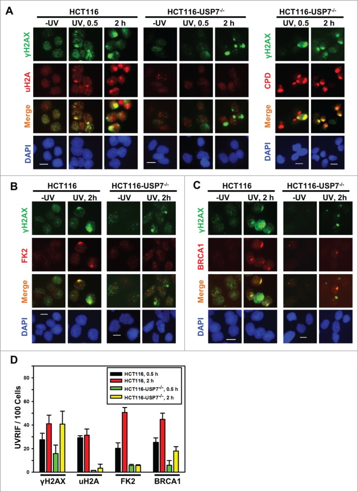
USP7 disruption compromises the formation of UVRIF of uH2A, FK2 and BRCA1 (A) HCT116 and HCT116-USP7−/− cells were exposed to micropore UV irradiation at 100 J/m2. Sub-nuclear spot accumulations of indicated DDR factors were visualized by immunofluorescence using specific antibodies. Calibration bar is 10 μm. Left panel: UVRIF of uH2A and γH2AX; right panel: UVRIF of CPD and γH2AX. (B) UVRIF of FK2 and γH2AX. (C) UVRIF of BRCA1 and γH2AX. (D) The quantitative data of γH2AX, uH2A, FK2 and BRCA1 foci from HCT116 and HCT116-USP7−/− cells. Mean ± SD were calculated from 4–6 microscopic fields of 3 independent experiments.