
Figure 5.
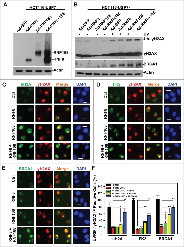
Adenovirus-mediated expression of RNF168 and RNF8 or168 partially rescues the formation of UVRIF of uH2A, FK2 and BRCA1 in HCT116-USP7−/− cells. (A) HCT116-USP7−/− cells were infected with the indicated adenoviral vectors expressing HA-tagged RNF8 and RNF168. Expression of RNF8 and RNF168 was examined by anti-HA Western blotting. (B) HCT116-USP7−/− cells were infected with indicated adenoviral vector or vector combination. The infected cells were harvested 2 h after UV exposure. The cell lysates from infected cells were examined by Western blotting for γH2AX and BRCA1 with anti-Actin blot as loading control. (C) The adenoviral vector infected cells were exposed to micropore UV irradiation at 100 J/m2. Two hour after UV irradiation, UVRIF of uH2A and γH2AX were visualized by immunofluorescence using specific antibodies. (D) UVRIF of FK2 and γH2AX. (E) UVRIF of BRCA1 and γH2AX. (F) Bar graph illustrates quantitative data of UVRIF. Mean ± SD of UVRIF vs. γH2AX positive cell ratio was calculated from 4–6 microscopic fields of 3 independent experiments. The p values were results from Student's t-test. Symbol * indicates P ≤ 0.05; Symbol ** indicates P ≤ 0.01. Calibration bar is 10 μm.