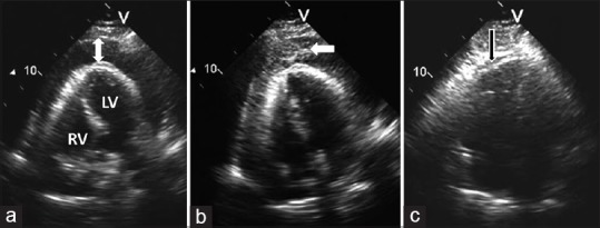
Figure 1.

Ultrasound-guided pericardiocentesis in a patient with malignant pericardial effusion and tamponade. (a) Apical view of the heart showing large circumferential pericardial effusion (arrow); (b) Intrapericardial injection of agitated saline (whitish-gray cloud of microbubbles of air) verifies correct positioning of the pericardiocentesis needle (arrow); and (c) following pericardiocentesis, the right ventricle has expanded and no residual pericardial effusion is seen within the pericardial sac (arrow). LV = left ventricle; RV = right ventricle
