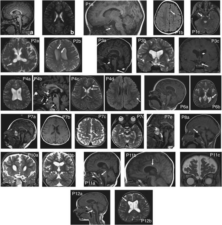
Figure 2.
Brain magnetic resonance imaging (MRI) findings – T1 and T2 images. (a, b): normal brain architecture of a 5-year old. MRIs obtained at the following ages: P1 36 m.o.; P2 18 m.o.; P3 10 y.o.; P4 4.2 y.o., P6 17 m.o.; P7 21 m.o.; P8 6 y.o.; P10 35 m.o.; P11 16 m.o.; P12 25 m.o. P1a – hypoplastic pituitary gland (HPG), P1b – gliosis, P1c – small brain stem (SBS) indicated by prominent prepontine cistern; P2a – large ventricles (LV), white matter hypomyelination (WMH), P2b – hypoplastic (thin) corpus callosum (HCC); P3a – SBS, thinning upper cervical cord, P3b – thin optic chiasm (TOC), P3c – LV, HPG; P4a – LV, P4b – TOC, HPG, SBS, P4c – frontal lobe atrophy (FLA), prominent frontal horns, P4d-WMH, gliosis; P6a – HCC, SBS, P6b – LV, WMH; P7a – microcephaly (MC), P7b – mild LV, P7c – WMH, P7d – SBS, P7e – SBS; P8a – MC, HPG; P10ab – LV, SBS, brain atrophy and WMH, gliosis in periventricular regions (not shown); P11a – HPG (arrowhead), SBS (white arrow), P11b – HCC, P11c – FLA; P12a – MC, SBS, HPG; P12b – LV, FLA. Original MRIs were reviewed by a UCLA Pediatric Neuroradiologist (NS).

