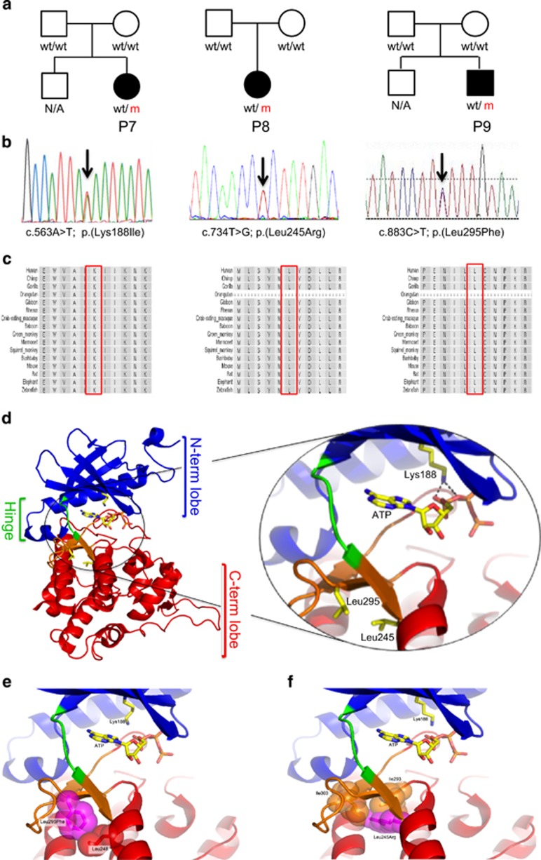
Figure 4.
Features of the three missense variants identified in DYRK1A. (a) The pedigree of three families with missense variants. (b) The Sanger sequencing traces of two of these variants. (c) Amino-acid alignments at the variant position (red box) and surrounding bases across 16 species as indicated on the left. (d) Overview of the human DYRK1A protein structure (PDB ID: 4MQ1). The active site (inset) is sandwiched between the N-terminal (blue) and C-terminal lobes (red), which are connected by a short hinge segment (green). The three residues mutated in patients (Lys188, Leu245, and Leu295) and the ATP (modeled from PDB ID: 1ATP) are shown as ball-and-stick configuration colored by atom. Lys188 coordinates (black dashed lines) the α- and β-phosphates of ATP. (e, f) Steric clashes caused by variants of Leu295Phe and Leu245Arg in relation to the active site. (e) Leu295Phe variant (magenta) causes a clash with Leu248 (red). (f) Leu245Arg (magenta) causes a clash with Ile303 (orange) and Ile293 (orange).

