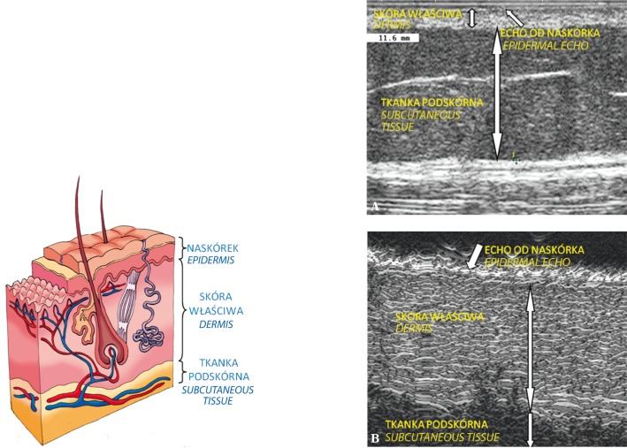
Fig. 5.
Ultrasound image of the skin with visible three layers: epidermal echo, dermis and subcutaneous tissue: A. image obtained by means of a classical scanner Philips HD11 XE with a linear-array transducer; B. scan obtained by means of a high-frequency ultrasound machine Episcan with a mechanical transducer of 50 MHz

