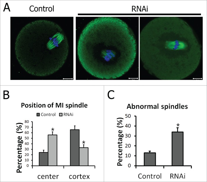
Figure 5.
Peripheral spindle migration at MI stage was impaired after TGN38 depletion in mouse oocytes. (A) Representative images of oocytes in different siRNAs injected groups. After cultured for 8.5 h following TGN38 RNAi, oocytes were fixed and co-stained with anti-α-tubulin (Green) antibody and Hoechst 33342 (Blue). Bars, 10 μm. (B) Percentages of position of MI spindle in oocytes. *, P < 0.05. (C) Percentages of oocytes with abnormal spindles. *, P < 0.05.

