Figure 1.
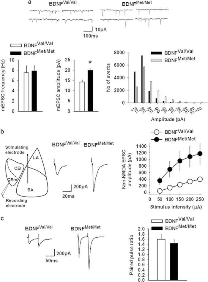
Non-NMDA receptor transmission is enhanced in the CEm neurons of BDNFMet/Met mice. (a) mEPSC frequency and amplitude in BDNFVal/Val (10 neurons/5 mice) and BDNFMet/Met (12 neurons/5 mice) groups. Upper panel shows examples of mEPSCs. Lower right panel shows event histogram for mEPSC amplitude. (b) EPSC amplitude in BDNFVal/Val (12 neurons/5 mice) and BDNFMet/Met (10 neurons/6 mice) groups. Left panel shows schematic presentation of the positions of stimulating and recording electrodes in the amygdala slice preparation. Middle panel shows examples of EPSCs evoked by 150 μA stimulation. (c) Paired pulse ratio in BDNFVal/Val (6 neurons/3 mice) and BDNFMet/Met (7 neurons/3 mice) groups. Left panel shows examples of EPSCs evoked at an interval of 60 ms. Asterisk denotes a statistically significant difference. BA, basal amygdala; CEl, latero-capsular subdivision of central amygdala; CEm, medial subdivision of central amygdala; LA, lateral amygdala.

