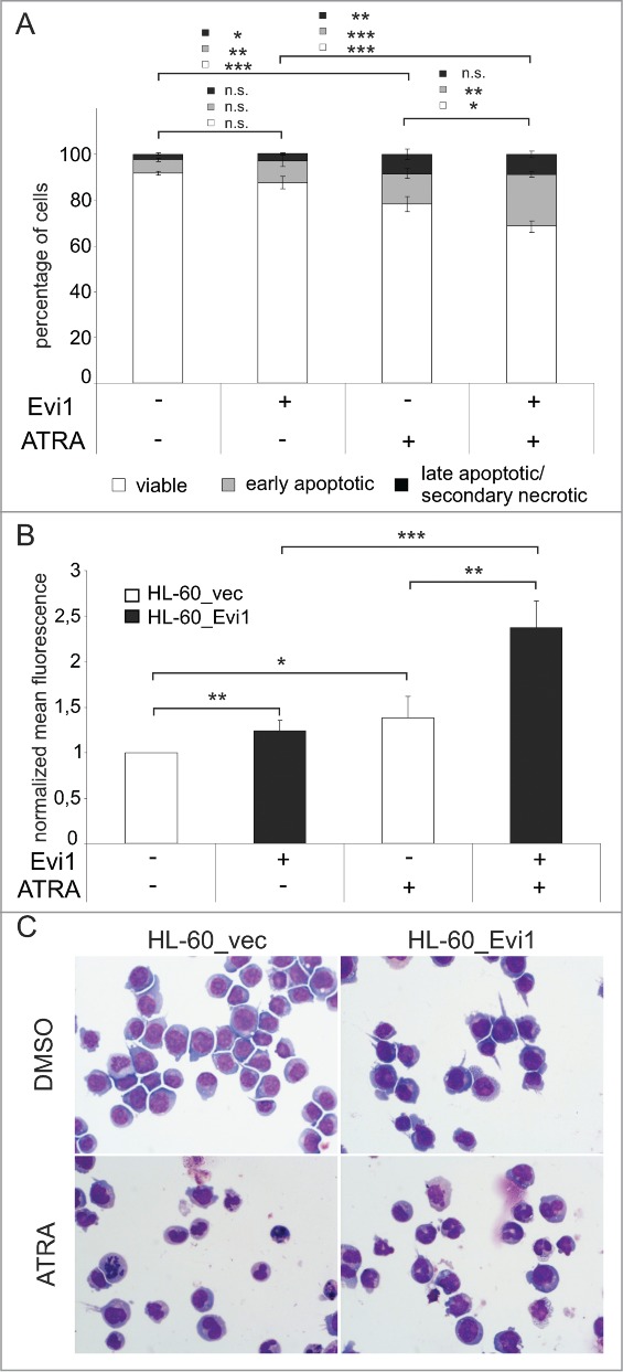
Figure 6.

Evi1 enhances ATRA induced apoptosis and differentiation in HL-60 cells. (A) HL-60_vec and HL-60_Evi1 cells were treated with solvent or ATRA for 5 days, stained with Annexin V and 7AAD, and analyzed by flow cytometry. Double negative cells were classified as viable (white portions of bars), Annexin V positive 7AAD negative cells as early apoptotic (gray portions of bars), and double positive cells as late apoptotic/secondary necrotic (black portions of bars).89 Results represent means +/- SEs of 8 independent experiments. (B) HL-60_vec cells (white bars) and HL-60_Evi1 cells (black bars) were treated with solvent or ATRA for 5 days, stained with PE-conjugated CD11b or isotype control antibodies, and subjected to flow cytometry. Results are expressed as the means of the PE fluorescence of CD11b stainings relative to that of the respective isotype controls and to untreated HL-60_vec cells, and represent the means + SEs of 7 independent experiments. Significance was calculated using Student's 2-tailed t-test (*, P < 0.05; **, P < 0.01; ***, P < 0.001). (C) Wright stained cytospin preparations of HL-60_vec and HL-60_Evi1 cells treated with solvent or ATRA for 5 d.
