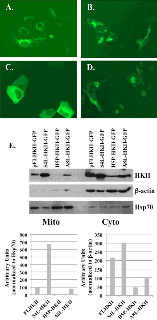
Figure 2. Fluorescent and immunoblot images of the HKII mutants with single point mutations.

U-2OS cells were transfected with plasmids carrying FLHKII or a point mutation of amino acids in position 4, 5, or 8. Stably-transfected cells were imaged at 400× magnification using an EVOS FL microscope. (A) U-2OS cells transfected with GFP-tagged wild-type FLHKII; (B) U-2OS cells transfected with (S4L)-HKII-GFP; (C) U-2OS cells transfected with (H5P)-HKII-GFP; (D) U-2OS cells transfected with (A8L)-HKII-GFP; (E) mitochondrial (Mito) and cytoplasmic (Cyto) fractions were isolated from U-2OS cells stably-transfected with wild-type FLHKII-GFP and each mutant HKII protein, then analysed by immunoblot. Immunoblots were probed with primary antibodies to detect: hexokinase II, β-actin and mtHsp70. Densitometry was used to quantify the expression levels of HKII in the cytoplasmic and mitochondrial cell fractions.
