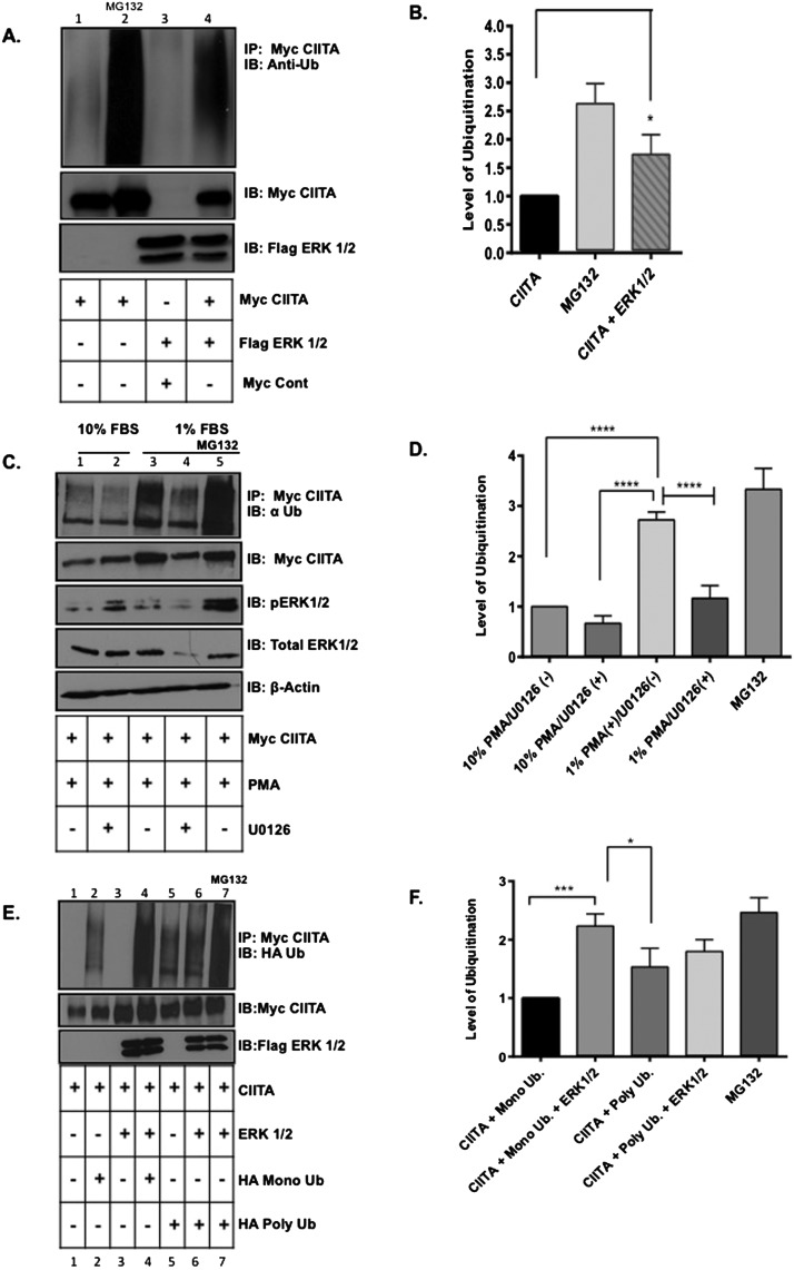
Figure 4. CIITA global ubiquitination and mono-ubiquitination is enhanced when ERK1/2 are overexpressed and inhibiting endogenous ERK1/2 leads to decreases in global CIITA ubiquitination levels.
In vivo ubiquitination assay: (A) COS cells were transfected with Myc–CIITA and Flag–ERK1/2. Lysate controls (bottom two panels) demonstrate expression of Myc–CIITA and Flag–ERK1/2. Data shown are cropped images from one IP gel and one lysate gel and are representative of three experiments. (B) Densitometry and quantification of data in Figure 4(A): Densitometry was performed on three independent experiments, ± S.D., *P<0.05. (C) COS cells were transfected with Myc–CIITA and indicated samples were treated with U0126 (MEK specific inhibitor) at time of transfections. Eighteen hours following transfections, 10% FBS media was replaced with 1% FBS media for 6 h and PMA and MG132 were added as indicated. Co-IP and ubiquitination analysis: Following all treatments, cells were harvested, lysed, pre-cleared and IP'd (immunoprecipitated) with anti-Myc antibodies. Lysate controls (bottom four panels) demonstrate expression of Myc–CIITA, total ERK1/2, phosphorylated ERK1/2 and actin as controls. Data shown are cropped images from one IP gel and one lysate gel. (D) Densitometry and quantification of data in Figure 4(C): Densitometry was performed on three independent experiments, ± S.D., ****P<0.0001. (E) COS cells were transfected with Myc–CIITA, Flag–ERK1/2, HA–mono-Ub or HA–poly-ubiquitin. MG132 was added to control sample for 4 h. Lysate controls (bottom two panels) demonstrate expression of Myc–CIITA and Flag–ERK1/2. Data shown are cropped images from one IP gel and one lysate gel. (F) Densitometry and quantification of data in Figure 4(E): Densitometry was performed on three independent experiments, ± S.D., ***P<0.001.

