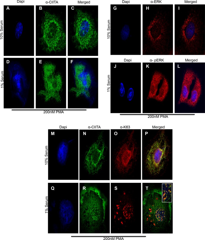
Figure 7. Lys63 Ub co-localizes with CIITA in cytoplasm and upon ERK1/2 activation moves to nucleus.
Immunofluorescence: COS cells were plated in six-well plates with glass coverslips. Twenty-four hours post seeding, cells were transfected with Myc–CIITA and HA–Lys63 ubiquitin. Eighteen hours following transfections, 10% serum media was removed where indicated and was replaced with 1% serum media for 6 h. Thirty minutes prior to harvest, 200 nM PMA was added to the indicated samples. Cells were fixed in ice-cold methanol for 10 min and were stained with anti-CIITA, anti-Lys63, anti-ERK1/2 and anti-pERK1/2 as indicated. (A–C) Cells in 10% serum media, stained for DAPI, CIITA and merge. (D–F) Cells in 1% serum free media, stained for DAPI, CIITA and merge. (G–I) Serum media (10%), stained for DAPI, ERK1/2 and merge. (J–L) Serum free media (1%), stained for DAPI, pERK1/2 and merge. (M–P) Serum media (10%), stained for DAPI, CIITA and Lys63 Ub and merge. (Q–T) Serum free media (1%), stained for DAPI, CIITA and Lys63 Ub and merge.

