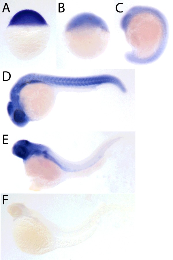
Figure 4. Spatiotemporal expression of naa10 revealed by in situ hybridization.

Lateral view of stained embryos at 0 hpf (A), 4 hpf (B), 18 hpf (C), 24 hpf (D) and 72 hpf (E). Negative control using the sense probe at 72 hpf (F).

Lateral view of stained embryos at 0 hpf (A), 4 hpf (B), 18 hpf (C), 24 hpf (D) and 72 hpf (E). Negative control using the sense probe at 72 hpf (F).