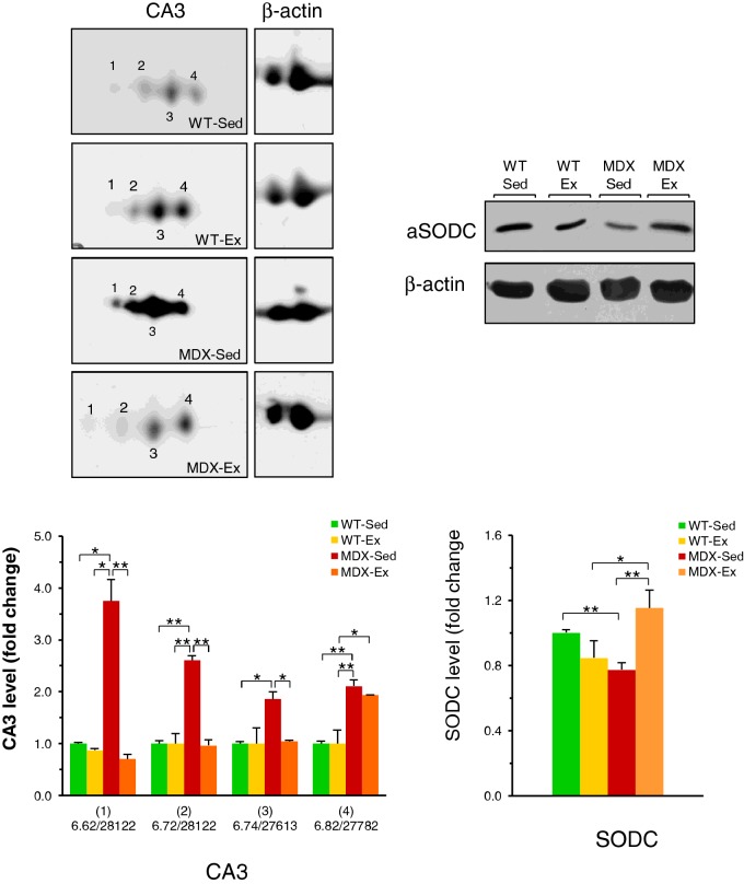
Figure 4. Western Blot of CA3 isoforms and SODC in quadriceps.
Representative immunoblots with expanded views of antibody-decorated protein spots and bands. Immunoblotting was performed after 2DE for CA3 and after 1DE for SODC. β-Actin was used as loading control. The blot densities are expressed as folds of control. CA3 isoforms are identified with pI-values and relative molecular masses: (1) pI 6.62/Mr 28122 Da; (2) pI 6.72/Mr 28122Da; (3) 6.74/Mr 27613 Da and (4) pI 6.82/Mr 27782 Da. Data are mean ± S.D.; *P<0.05; **P<0.01. Sedentary WT (WT-Sed), exercised WT (WT-Ex), sedentary mdx (MDX-Sed) and exercised mdx (MDX-Ex) mice.

