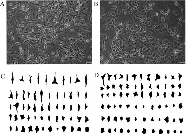
Figure 2. Images of cell shape in segment pre-and post.
(A) An image with points of grid stimulated by TGF-β1. (B) An image with points of grid never stimulated by TGF-β1. (C) The image stimulated by TGF-β1 with segmented and sorted cells. (D) The image stimulated never by TGF-β1, with segmented and sorted cells. The images were captured by phase contrast microscope (Olympus CKX41-A32PH, Tokyo, Japan) with a LCACHN20XPH 20×/0.4 NA objective (bars, 20 μm).

