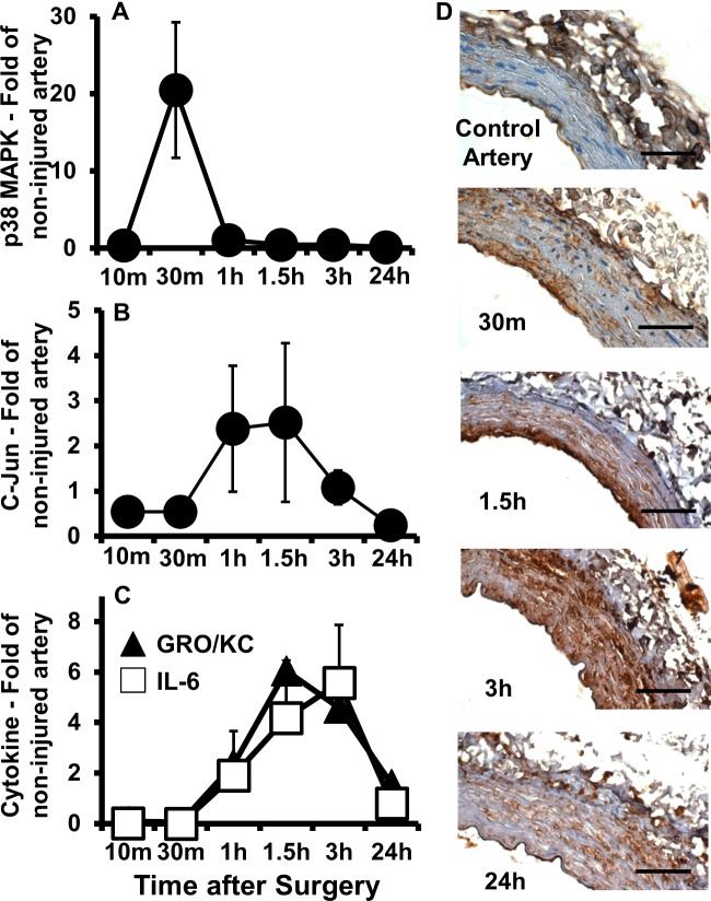
Figure 3. Quick activation of MAPK pathways and temporal accumulation of vascular cytokines in injured compared with control arteries.
(A–C) Levels of phosphorylated p38 MAPK (A), phosphorylated c-Jun (B) and the inflammatory cytokines IL-6 and GRO/KC (C) over time in injured compared with non-injured contralateral arteries as determined using multiplex immunoassays. Each point represents the mean ± S.E.M. (n=5). (D) Representative immunohistochemistry cross sections stained for IL-6 in control and injured arteries harvested at 30 min, 1.5 h, 3 h and 24 h after balloon injury. Scale bar=50 μm.

