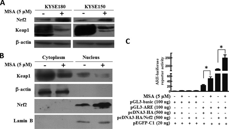
Figure 1. MSA activated Keap1/Nrf2 pathway in ESCC cells.
(A) KYSE180 and KYSE150 were treated with MSA (5 μM) for 24 h. Then, total cell lysates were prepared. Western blotting was performed to examine Keap1 and Nrf2. β-Actin was used as a loading control. (B) KYSE150 cells were treated with MSA (5 μM) for 24 h. Then, protein lysates were prepared. Keap1 and Nrf2 were detected in the cytoplasmic and nuclear extracts by western blot. β-Actin was used as a loading control of cytoplasmic protein and lamin B was shown as a loading control of nuclear protein. (C) KYSE150 cells were transfected with 100 ng of pGL3-ARE or pGL3-basic and 500 ng of pcDNA3-HA or pcDNA3-HA/Nrf2 plasmids with or without MSA treatment respectively. The pEGFP-C1 (20 ng) plasmid was co-transfected to normalize transfection efficiency. The luciferase activities were measured 48 h after transfection. Means ± S.D., n=3, *P<0.05.

