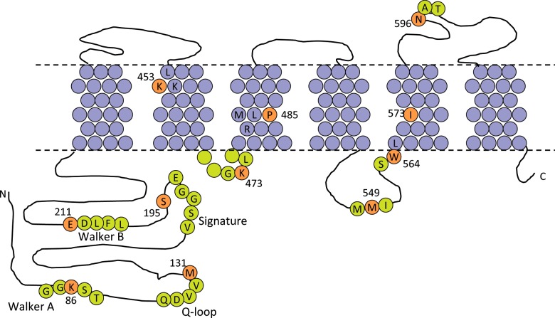
Figure 1. Location of the residues selected for mutation in ABCG2.
The transmembrane topology of ABCG2 is represented according to the data of Mao and colleagues [30], with approximate membrane plane indicated by the dashed line. TM residues are shaded purple and extramembraneous residues in lime. Residues for mutation are on an orange background, numbered and indicated using the single-letter amino acid code. Local sequence context and NBD sequence motifs have been shown for ease for reference.

