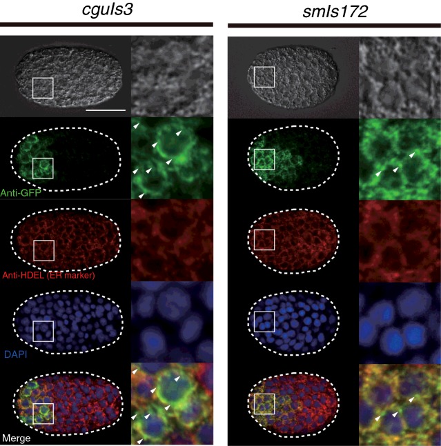
Figure 5. Characterization of the two NUC-1 fusion proteins in transgenic animals of smIs172 and cguIs3.
The subcellular localization of NUC-1 fusion proteins in smIs172 and cguIs3 embryos. Immunostaining with antibodies against GFP (in 1:500 dilution) and against HDEL (ER marker, in 1:200 dilution) were performed on embryos of smIs172 and cguIs3. Images are indicated with conditions of microscopy for photography. Nucleus was labelled with DAPI. The anterior of embryo is shown toward left and those cells located in the anterior region are enlarged to show co-localization of NUC-1 fusion proteins with ER proteins portion in detail. Imperfect merged of GFP and ER markers are indicated by arrows. Scale bar: 20 μm.

