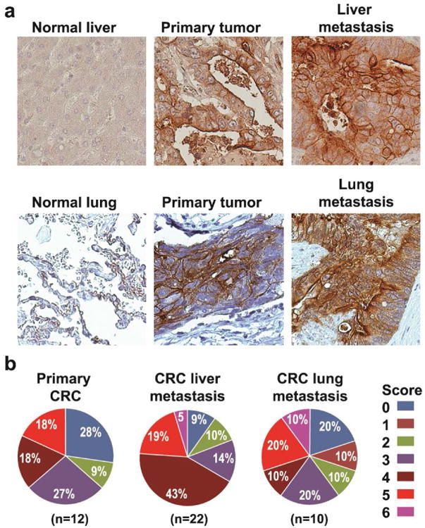
Figure 2. Analysis of FRα expression in CRC liver and lung metastasis.

a Examples of immunohistochemical staining for FRα in liver and lung CRC metastases. Positive FRα staining of CRC was cytoplasmic or membranous or both; most positive cases showed both patterns. FRα staining was negative in normal liver and lung tissues. b. Differences in proportion of positive cells and intensity of staining were noted in positively stained cases and formed the basis of our grading system. Comprehensive total score that weighs both factors was calculated by summation of proportion and intensity values. High FRα expression (score 3-6) was detected in 63% of primary CRCs, 81% of CRC liver metastases and 60% of CRC lung metastasis.
