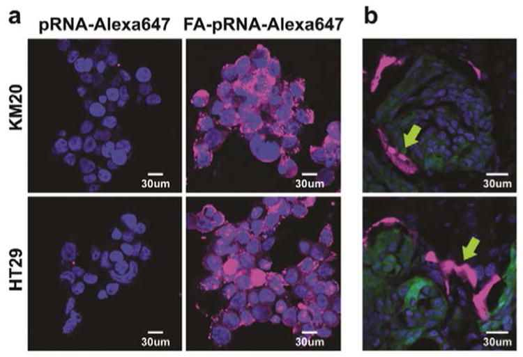
Figure 3. FA-pRNA nanoparticles binding to CRC cells.

a. Binding and entry of FA-pRNA-Alexa647 nanoparticles into KM20 and HT29 cells in vitro. Magnification 40×. b. Single dose (4μg/g in 100 μl of PBS) of FA-pRNA-Alexa647 labeled nanoparticles was administered intravenously into mice with HT29 liver metastases. Accumulation of fluorescently-labeled nanoparticles was evaluated microscopically 2h after RNA nanoparticle administration. Yellow arrow: pRNA-Alexa647 (top panel), FA-pRNA-Alexa647 (bottom panel); green: GFP-expressing cancer cells; blue: DAPI stain for nuclear dsDNA; magenta: Alexa647. Magnification 40×.
