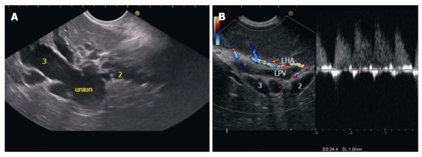
Figure 3.

Union of segmental ducts. A: Segment 2 is identified as duct coming from cranial part of liver segment and segment 3 duct is identified as duct coming from caudal part of liver segment; B: Sometimes the ducts are not dilated and in such situation the tributaries of left portal vein can be identified after application of color doppler and followed to the union and formation of umbilical part of portal vein. LPV: Left portal vein; LHA: Left hepatic artery.
