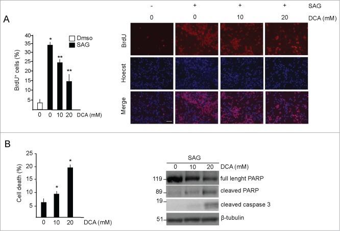
Figure 2.
DCA decreases proliferation and induces apoptosis in GCPs. (A) Left, DCA decreases proliferation in GCPs. BrdU incorporation assay in cells treated with DMSO or with SAG (200 nM, 48 hours) and increasing amounts of DCA for 48 hours. BrdU was added for the last 24 hours. Right, representative images of BrdU staining. Scale bar = 50 μm. *SAG vs control, P < 0.05; **DCA versus SAG, P < 0.01. (B) Left, DCA increases cell death in SAG-treated GCPs, as measured by the increase of the number of condensed and fragmented nuclei compared to control. Right, DCA increases active PARP and caspase 3. *DCA versus control, P < 0.01; n = 4.

