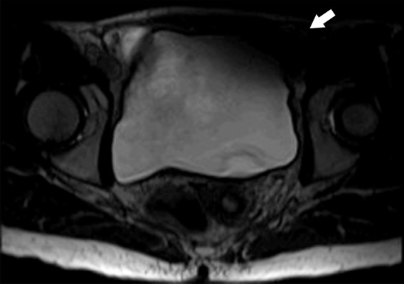
Figure 18b.

Parallel imaging artifact caused by a missing multichannel receive coil in a 47-year-old woman undergoing evaluation for uterine fibroids. (a) Coronal T2-weighted MR image shows placement of the parallel imaging receive coil array over the patient. The large light-gray circle represents B0, and multichannel receive coils (small dark-gray circles) overlie the pelvis. One of the lower coil elements is out (black circle). (b) Axial T2-weighted MR image shows marked signal loss (arrow) in the left anterior portion of the pelvis.
