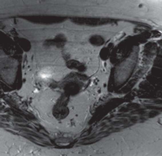
Figure 21b.

Aliasing artifact along the section direction in a three-dimensional acquisition. (a) Axial T2-weighted MR image acquired with a three-dimensional fast spin-echo sequence (SPACE [Sampling Perfection with Application optimized Contrasts using different flip angle Evolutions]; Siemens Healthcare) shows an artifact from the perineum (arrows) overlying the upper portion of the pelvis. (b) Repeat axial T2-weighted fast spin-echo (SPACE) MR image with oversampling in the section direction shows amelioration of the artifact at the cost of increased imaging time. (c) Coronal T2-weighted MR image shows the imaged FOV (red rectangle) and the oversampled volume (green dashed rectangle).
