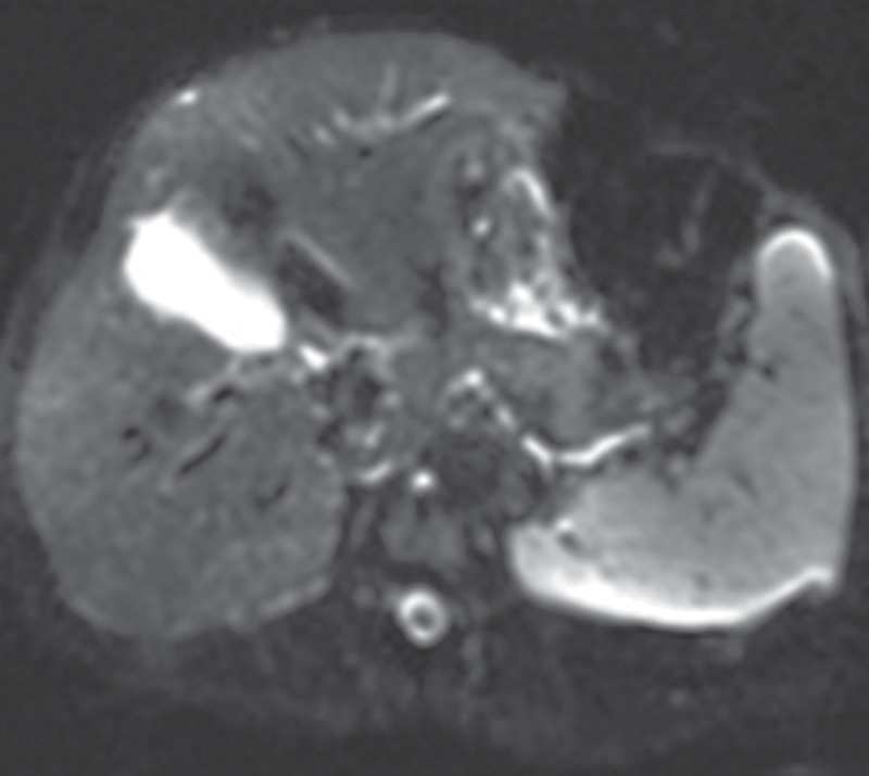
Figure 6c.

Echo-planar imaging artifacts at diffusion-weighted MR imaging. (a) Coronal whole-body contrast-enhanced MR image shows the B0 field (green dashed rectangle) and the excitation slab (red rectangle) for echo-planar diffusion-weighted imaging. (b) Axial diffusion-weighted MR image of the upper part of the abdomen shows the effect of B0 inhomogeneity over the lungs, which, in combination with poor fat suppression and eddy current effects, leads to noticeable phase-encoding artifact. (c) Axial diffusion-weighted MR image of the upper part of the abdomen obtained with improved shimming and with fat suppression shows decreased image distortion and better image quality.
