
Figure 7b.
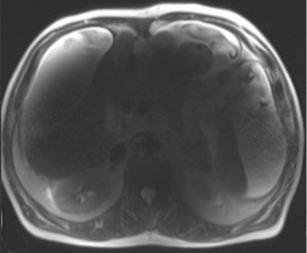
B1 inhomogeneity (standing waves) at 3-T MR imaging of a patient with cirrhosis and ascites. Coronal T2-weighted (a) and axial out-of-phase T1-weighted (b) MR images show a signal void in the center of the images because the wavelength of the RF transmission field is on the same order of magnitude as the dimension of the patient. The resulting variations in the RF transmission field produce focal areas of decreased signal intensity.