
Figure 5:
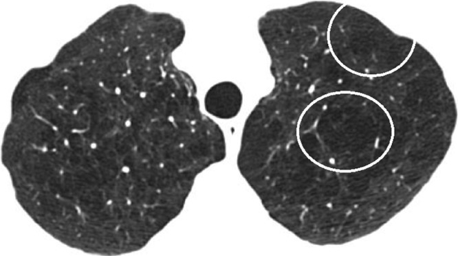
Confluent CLE. CT scan in patient with GOLD stage I COPD shows multiple lucencies that span several secondary pulmonary lobules (circled in left lung) but are not associated with extensive hyperexpansion of secondary pulmonary lobules or distortion of pulmonary architecture.