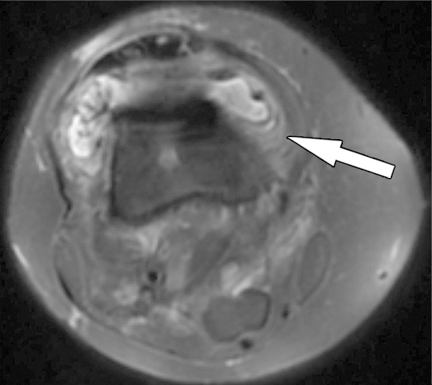
Figure 15b.

Periprosthetic joint infection after total knee arthroplasty in a 60-year-old man. Axial (a) and sagittal (b) intermediate-weighted fast SE and axial STIR (c) MR images demonstrate thickening, layering or lamellation, and signal hyperintensity of the synovium (arrow) with expansion of the joint capsule. Joint aspiration and microbiological analysis resulted in identification of Staphylococcus aureus bacteria.
