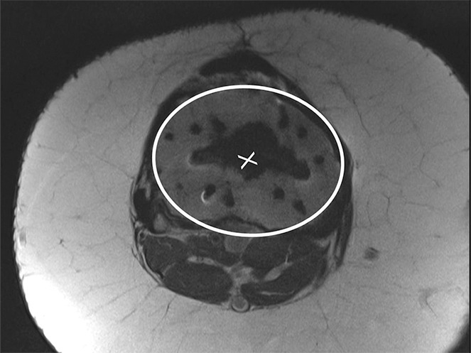
Figure 17b.

Determination of the rotational angle of the tibial component after total knee arthroplasty in a 63-year-old woman. (a) Axial intermediate-weighted fast SE image demonstrates the tibial component anteroposterior axis, which intersects the center of the anterior and posterior margins of the tibial component. (b) Axial intermediate-weighted fast SE image demonstrates determination of the geometric center of the tibia plateau (x) by a best-fit ellipse (solid line) circumscribing the tibial cortex. (c) Axial intermediate-weighted fast SE image demonstrates the tibial tubercle axis (solid line) as the line connecting the determined geometric center of the tibia (x in b–d) through the center (arrow) of the tibial tubercle. (d) Axial intermediate-weighted fast SE image demonstrates the rotational angle of the tibial component measured as the angle between the tibial tubercle axis (solid line in c and d) and tibial component axis (dashed line in a and d).
