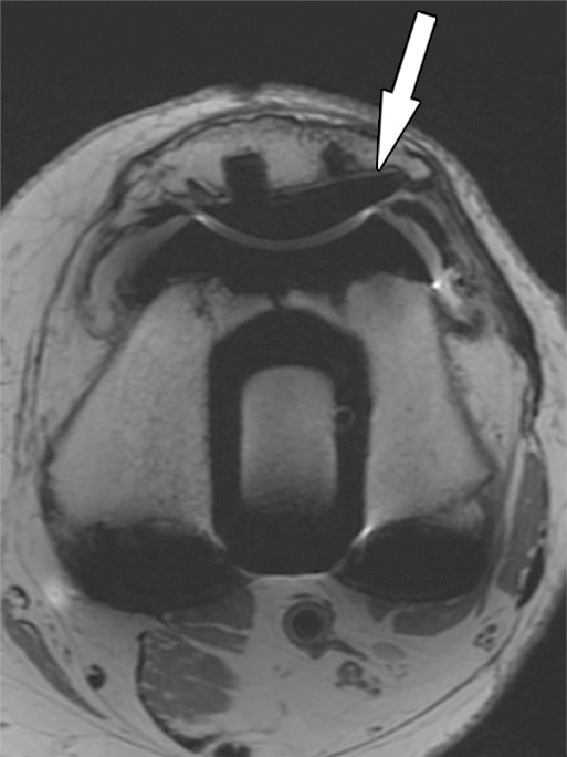
Figure 4b.

Fibrous membrane formation after total knee arthroplasty in a 70-year-old man. Axial intermediate-weighted fast SE images show a thin layer of increased signal intensity at the implant-bone interface of the tibial (arrow in a) and patellar (arrow in b) components, which is surrounded by a thin layer of decreased signal intensity.
