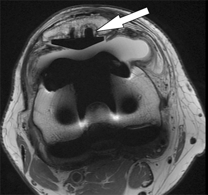
Figure 5a.

Periprosthetic bone resorption after total knee arthroplasty in a 77-year-old woman. Axial (a) and sagittal (b) intermediate-weighted fast SE MR images demonstrate bone resorption at the implant-cement interfaces of the patellar (arrow in a), femoral (white arrow in b), and tibial (black arrow in b) components, seen as irregular layers of increased signal intensity, which are surrounded by a layer of decreased signal intensity. Additionally, there is synovitis visible on the images.
