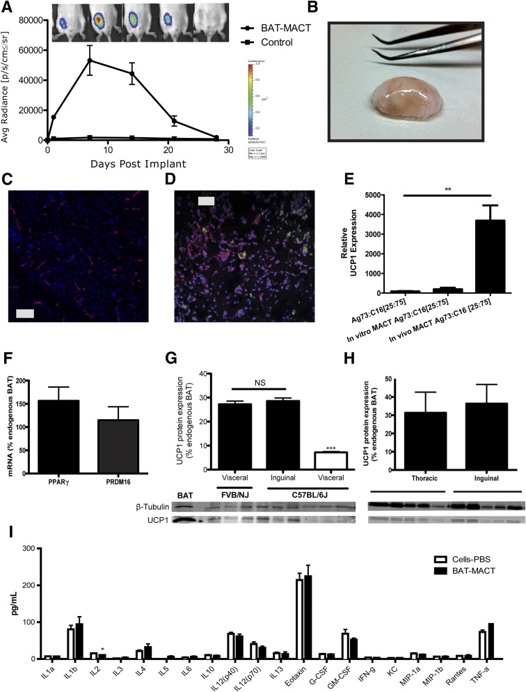
Figure 3.
BAT-MACT in vivo characteristics. A: Persistence of BAT-MACT over time monitored by luciferase activity in live animals (FVB/NJ; n = 5); false color heat scale image indicating average (Avg) photon radiance. Max, maximum; Min, minimum; sr, steradian. B: Macroscopic morphology of implants after 2 weeks. BAT-MACTs were removed after 14 days; fixed, cryosectioned, and stained with DAPI; and stained for the vascularization marker endomucin (C) or neutral lipids (green) and UCP1 (red) (D). E: mRNA expression of UCP1 relative to WAT using samples from differentiated ADMSCs plated on Ag73 and C16 peptide-coated TCPS or in Ag73- and C16-conjugated AcHyA hydrogels cultured in vitro or implanted into recipient animals (FVB/NJ; n = 4, 3, or 6, respectively). F: PRDM16 and PPARγ mRNA expression normalized to GAPDH of BAT-MACTs after 2 weeks in vivo relative to endogenous BAT (FVB/NJ; n = 9). G: UCP1 protein expression of BAT-MACTs generated with ADMSCs from visceral or subcutaneous FVB/NJ or C57BL/6J mice 2 weeks postimplantation into a syngenic recipient relative to endogenous BAT (n = 3). NS, not significant. H: UCP1 protein expressed in BAT-MACTs of different implant sites after 2 weeks in vivo relative to endogenous BAT (FVB/NJ; n = 5). I: Serum cytokine levels measured 2 weeks postimplantation of BAT-MACT or the same ADMSCs delivered via PBS. Animals were administered the 60% fat diet for the duration of this study (C57BL/6J; n = 6). IL, interleukin. Significance at **P < 0.01 and ***P < 0.001.

