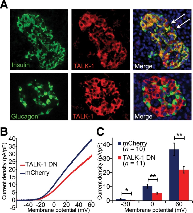
Figure 2.
Human β-cells contain functional TALK-1 channels. A: Immunofluorescence staining of TALK-1 (red) and insulin (green, upper panels) or glucagon (green, lower panels) in human pancreas sections; nuclei (blue) are shown in the merge panel. White arrows indicate TALK-1–positive, insulin-negative cells. B: K2P current obtained in human β-cells expressing either control mCherry or TALK-1 G110E P2A mCherry in response to a voltage ramp from −120 mV to 60 mV; currents are plotted between −60 and 60 mV. C: Quantification of current densities at indicated membrane potentials. Data are mean ± SEM. *P < 0.05; **P < 0.005.

