
Figure 4.
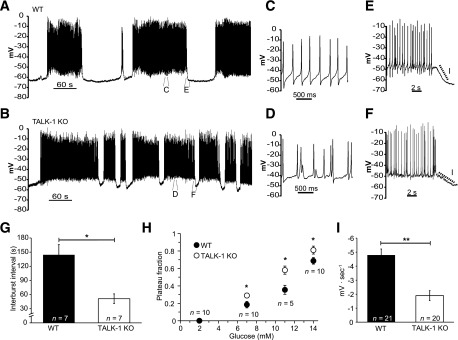
TALK-1 regulates β-cell electrical activity. A: Representative Vm recording from a WT β-cell recorded in an intact mouse islet stimulated with 14 mmol/L glucose. B: Typical Vm recording from a TALK-1 KO β-cell in an intact islet in the presence of 14 mmol/L glucose. C and D: Enlarged view of APs recorded from WT (C) and TALK-1 KO (D) β-cells in 14 mmol/L glucose. E and F: Enlarged view showing the slope of Vm repolarization at the termination of an electrical oscillation in WT (E) and TALK-1 KO (F) β-cells. G: Average length of the electrically silent interburst interval in WT and TALK-1–deficient islets, which was measured during the first 20 min of electrical excitability induced with 14 mmol/L glucose. H: Plateau fraction of electrical excitability in islets, determined as in G. I: Mean slope of Vm repolarization at the termination of each oscillation of electrical activity in WT and TALK-1 KO β-cells, which was measured at the end of each oscillation in electrical excitability as in G. Data are mean ± SEM. *P < 0.05; **P < 0.005.