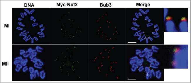
Figure 2.

The localization of Bub3 and Myc6-Nuf2 on chromosome spreads at MI and MII stages. Oocytes were injected with a low concentration of Myc6-Nuf2 mRNA and cultured to MI and MII stages, then chromosomes were spread and stained with anti-Myc6 antibody (green), anti-Bub3 antibody (red) and Hoechst 33342 (blue). Magnifications of the boxed regions are shown on the right of each main panel. Scale bars: 10 µm.
