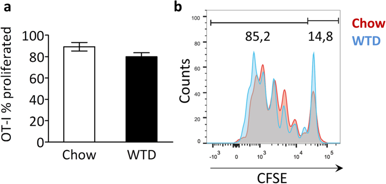
Figure 2. Cross-presentation occurs under hyperlipidemic conditions.

Ldlr−/− mice (n = 3) on a normal chow diet or fed a Western type diet (WTD) for three weeks were iv injected with irradiated OVA-expressing splenocytes and CFSE-labeled OT-I Tcells. After 72 hrs, spleens were harvested and cross-presentation was assessed by flow cytometry, quantifying the proportion of proliferating OT-I Tcells (cells with a diluted CFSE signal) within the total OT-I Tcell population, normalized for amount of injected cells. (a) Bar graph of proliferated OT-I Tcells (% of total OT-I Tcells) in spleen of chow or WTD-fed ldlr−/− mice. (b) Representive CFSE dilution peaks of the OT-I Tcell population. Data are presented as mean ± SEM.
