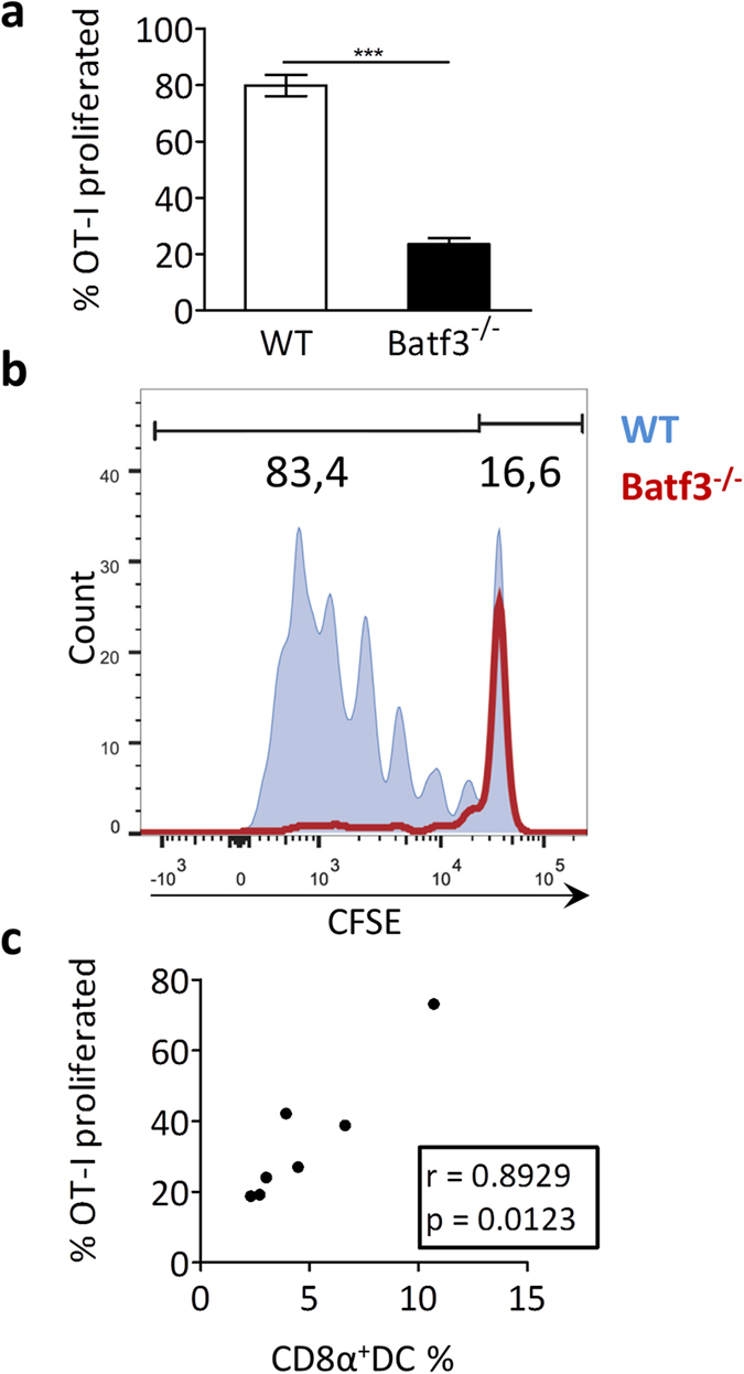
Figure 4. Cross-presentation is affected in batf3−/− chimeric mice.

Batf3−/− chimeric or wt ldlr−/− mice (n = 7) were iv injected with necrotic OVA-expressing splenocytes and CFSE-labeled OT-I T cells. After 72 hrs, spleens were harvested and cross-presentation was assessed by flow cytometry, quantifying the proportion of proliferating OT-I Tcells (cells with a diluted CFSE signal) within the total OT-I Tcell population, normalized for amount of injected cells. (a) Bar graph of proliferated OT-I Tcells (% of total OT-I Tcells) in spleen. (b) Representive CFSE dilution peaks of the OT-I Tcell population. (c) Correlation analysis between amount of residual CD8α+ DCs and the remaining cross-presentation capacity in batf3−/− chimeras. Data are presented as mean ± SEM, ***p < 0,001.
