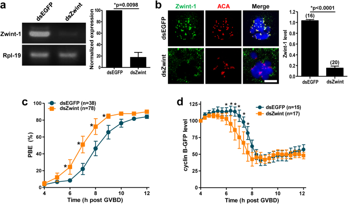
Figure 1. Zwint-1 knockdown accelerates polar body extrusion.
GV oocytes injected with Zwint-1 dsRNA (dsZwint) were cultured for 13 h in the presence of IBMX, washed in IBMX-free medium, and allowed to progress to MetI. (a) RT-PCR of Zwint-1-depleted oocytes. Rpl-19 was used as a loading control. Normalized expression of Zwint-1 was quantified from two independent experiments and shown to the right panel (*p = 0.0098). (b) Immunostaining of Zwint-1-knockdown oocytes. Oocytes were fixed at 6–7 h after GVBD and immunostained with anti-Zwint-1 antibody. Kinetochores and DNA were stained with anti-centromere antibody (ACA) and DAPI, respectively. Shown are representative of three independent experiments with at least 30 oocytes. Scale bar, 10 μm. Quantification of Zwint-1 signals is shown in the right of images (*p < 0.0001). The number of oocytes analyzed is shown above the bars. (c) Timing of polar body extrusion (PBE) was determined in Zwint-1-knockdown oocytes. Data are mean ± SEM from three independent experiments with the indicated number of oocytes (*p < 0.05). The mean time at which half of oocytes completed PBE was 6 h 58 min for Zwint-1 knockdown and 8 h 5 min for control. (d) Oocytes depleted of Zwint-1 were injected with mRNA encoding cyclin B-GFP, and GFP levels were measured every 20 min up to 8 h. Quantification of cyclin B levels is shown. For each oocyte, the fluorescence intensity was normalized to the intensity recorded 4 h after GVBD. Data are mean ± SEM from one experiment with the indicated number of oocytes (*p < 0.05). Note that the cyclin B degradation was initiated around 7 h after GVBD for control and 6 h after GVBD for Zwint-1-knockdown oocytes.

