
Figure 6.
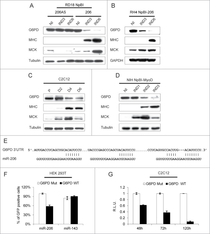
G6PD is downregulated during myogenic differentiation and is a direct target of miR-206. (A) Western blot analysis of the indicated proteins in RD18 cells infected with either a control (NpBI-206AS, antisense) or a miR-206-expressing (NpBI-206) lentiviral vector, treated or not with doxycycline for the indicated days (not induced, NI; induced, IND). (B) Western blot analysis of the indicated proteins in RH4 NpBI-206 cells, treated or not with doxycycline for the indicated days (not induced, NI; induced, IND). (C) Western blot analysis of the indicated proteins in C2C12 cells grown in proliferation medium (P) and after 2, 4 and 6 days in differentiation medium (D2, D4 and D6). (D) Western blot analysis of the indicated proteins in NIH NpBI-MyoD cells treated or not with doxycycline for the indicated days (MyoD not induced, NI; MyoD induced, IND). (E) Schematic representation of the 3 MREs of the human G6PD 3’ UTR aligned with the miR-206 sequence. The complementary of the miR-206 sequence with the MREs is indicated. (F) Flow cytometry quantification of GFP expression in HEK 293T cells co-transfected with wild type or mutant G6PD sensor construct along with miR-206 or miR-143 as a control. (G) Relative luciferase expression of C2C12 cells transfected with either wild type or mutant G6PD luciferase sensor construct, after 48, 72 and 120 hours in differentiation medium. For each time point, luciferase counts obtained with the control plasmid were set at 1.