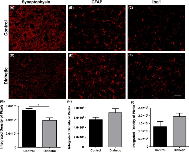
Figure 2.

Immunohistochemistry at lamina IX. (A, D) Synaptophysin. (B, E) GFAP (glial fibrillary acidic protein). (C, F) Iba1 (ionized calcium‐binding adapter molecule 1). (G–I) Quantification of the integrated density of pixels of synaptophysin, GFAP, and Iba1, respectively. *P < 0.05.
