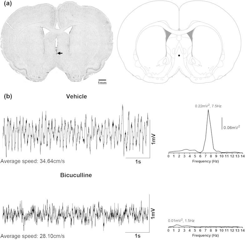
Figure 1. Microinjection of bicuculline into the medial septum attenuates hippocampal theta wave activity in the awake rat during exploration in open field.
(a) Digital image of a Nissl-stained coronal section (left panel) and its corresponding diagrammatic representation (right panel). The coronal section is taken through medial septum and shows the microinjection site which is given by the dye spot (arrow). The diagrammatic representation is adapted from Paxinos and Watson53. The filled circle on the diagrammatic representation corresponds to the microinjection site on the digital image and was judged to be within the medial septum. (b) are the representative waveforms of hippocampal field activity recorded from stratum radiatum of the hippocampus. The waveforms were recorded during exploration in the first minute following placement of the animal into the open field chamber. The average speed of ambulation in 10-s period around the trace is given underneath each trace. Animals were microinjected with vehicle (top; 0.5 μl) or bicuculline (bottom; 0.125 μg/0.5 μl) into the medial septal region immediately prior to the start of the open field experiment. Clear theta wave activity was observed in the vehicle-injected animal and, in line with visual observation, the peak frequency of the associated Fast Fourier Transform (FFT; frequency resolution 0.5 Hz; on the right of each trace) of the hippocampal field activity was in the theta range (4–12 Hz) and was clearly demarcated from other frequencies around the region of the peak. However, microinjection of bicuculline resulted in a loss of theta wave activity and FFT analyses revealed a peak in the non-theta range.

