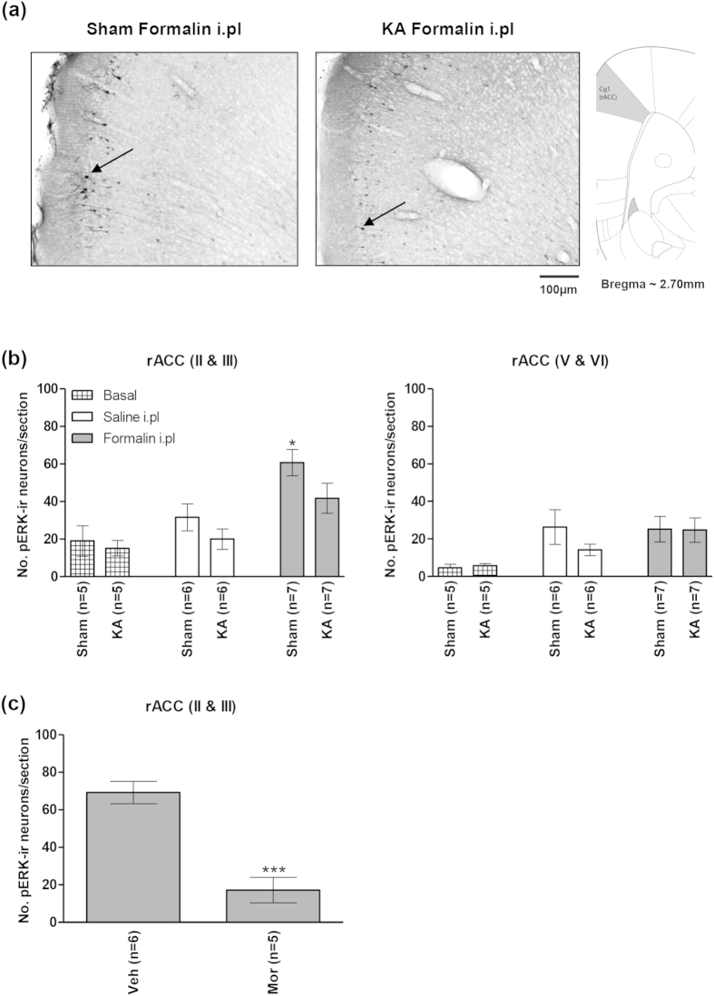
Figure 7. Kainic acid-induced lesion of the medial septal region blocked formalin-induced increase in pERK in the rostral anterior cingulate cortex.
At the top are digital representations of sections through the rostral anterior cingulate cortex (rACC; (a)). The digital panels show pERK-immunoreactive (pERK-ir) neurons (e.g. those next to arrows in (a)) observed on intraplantar (i.pl) injection of 2.5% formalin (‘Formalin i.pl’) into right hind paw of ‘KA’ and ‘Sham’ animals. The ‘KA’ and ‘Sham’ animals were pre-treated with microinjection of kainic acid and vehicle, respectively, into the medial septal region. The effect of KA pre-treatment on populations of neurons in the MSDB is shown in Fig. 4. Note that in the ‘Sham’ group (b), hind paw injection of formalin, but not saline (‘Saline i.pl’) evoked a significant increase in number of pERK-ir neurons in layers II and III of rACC that was attenuated with KA pre-treatment ((b), left). Hind paw injection of formalin did not evoke a significant increase in the expression of pERK-ir in rACC layers V and VI of the ‘Sham’ group ((b) right). In (c), pre-treatment with intraperitoneal (i.p) morphine (5 mg/kg, Mor) significantly reduced the formalin-induced pERK-ir in rACC layers II and III as compared to saline-injected controls (Veh). Data are mean ± SEM. Statistical significance: (b) rACC (II & III) *p < 0.05 vs. all other groups; (c) rACC (II & III) ***p < 0.001 vs. Veh. Statistical analyses were performed using one-way ANOVA (b) or unpaired t-test (c).

