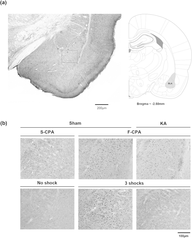
Figure 9. Egr-1/Zif268 was induced in the basolateral amygdala during conditioned place avoidance and contextual fear.
((a) left panel) is digital image of a Egr-1/Zif268-immunolabelled coronal section taken through the basolateral amygdala (BLA). The corresponding diagrammatic representation and the related anterior-posterior coordinate are on right (a). The diagrammatic representation is adapted from Paxinos and Watson53. (b) are digital images showing Egr-1/Zif268-immunoreactivity (ir) in the area of BLA that corresponds to the boxed region shown in (a). Nuclei of cells containing Egr-1/Zif268-ir stand out darkly stained relative to the background on the digital images. The sections in (b) are taken from ‘Sham’ and ‘KA’ animals that were pre-treated with microinjection of vehicle and kainic acid, respectively. The sections are from animals used to develop Fig. 10. The effect of KA pre-treatment on population of neurons in the MSDB is shown in Fig. 4. The treated animals were either (i) conditioned with saline-saline (‘S-CPA’ or saline-conditioned place avoidance) or saline-formalin (‘F-CPA’ or formalin-conditioned place avoidance), or (ii) conditioned with footshocks (‘3 shocks’) in the contextual fear task. Animals that did not receive any footshock (‘No shock’ on figure) acted as controls for the ‘3 shocks’ group. Notice the relatively high number of Egr-1/Zif268-ir cells in the sections taken from ‘Sham F-CPA’ and ‘Sham 3 Shocks’ animals ((b) middle panels) as compared to the corresponding control ((b) left panels). On the other hand, little or no Egr-1/Zif268-ir was observed in ‘3 shocks’ animal following pre-treatment with KA ((b) lower row, right panel).

