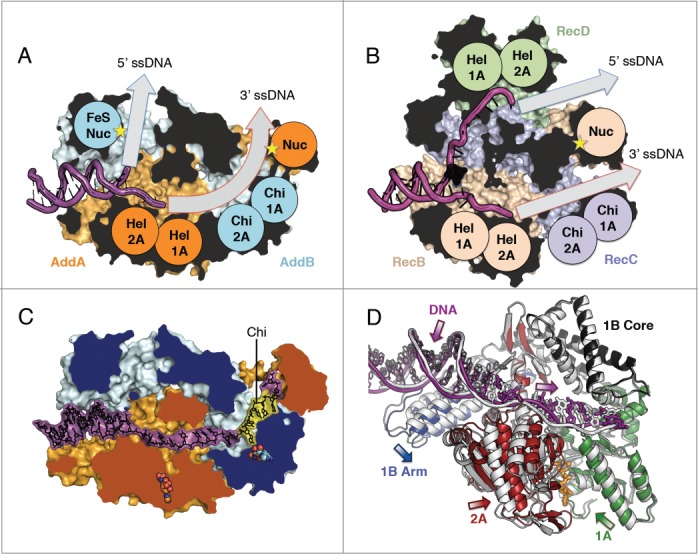
Figure 1.

AddAB and RecBCD structural architectures. Clipped surface view of (A) AddAB initiation complex (pdb code 3u44) and (B) RecBCD initiation complex (pdb code 3k70) with helicase motor (Hel), Chi-scanning and nuclease (Nuc) domains labelled and respective ssDNA tunnels highlighted (grey arrows). The clipped interior surface is coloured black. (C) AddAB structure in complex with Chi (yellow) (pdb code 4cej) with 2 bound ADPNP molecules shown as spheres (orange AddA and cyan AddB respectively). The clipped surface is coloured by protein. (D) Translocation-like movements of AddA helicase domains upon binding of ADPNP. Overlay of the translocation-like binary (grey) (pdb code 4ceh) and ternary ADPNP-bound (coloured by domain) (pdb code 4cei) AddAB structures showing relative displacement of the helicase motor and 1B arm domains. The single-stranded portion of DNA (thick bonds) moves away from the junction, corresponding to the closing movement of the 2A domain, whilst movement of the duplex (thin bonds) is coupled to the arm domain. Bound ADPNP is shown in sticks with the Mg2+ ion as a sphere (orange). The structures were aligned based on the 1B core domain and the 2B domain, which is hidden for clarity.
