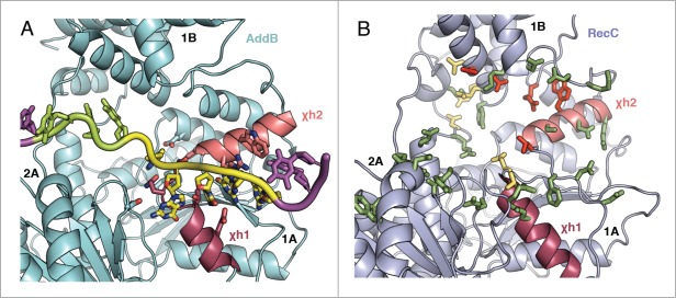
Figure 5.

Comparison of the positions of Chi-interacting residues in AddB and RecC. The Chi-binding site of AddB from the Chi-bound crystal structure (pdb code 4cej) is summarised (A) with key residues shown with sticks. Alongside is the same region of RecC (B) with mutations from the work by Handa et al.42 shown as sticks. Mutations that didn't affect Chi recognition are shown in green, Type I mutations that abolished Chi-binding are in red and Type II mutations that increased the promiscuity of Chi-sequence recognition are in yellow-orange. The 2 Chi-helices χh1 and χh2 are coloured similarly in each structure.
